Effects of Resistance Training and Bowdichia virgilioides Hydroethanolic Extract on Oxidative Stress Markers in Rats Submitted to Peripheral Nerve Injury


 ,
,  ,
,  and
and
Abstract
:1. Introduction
2. Methods
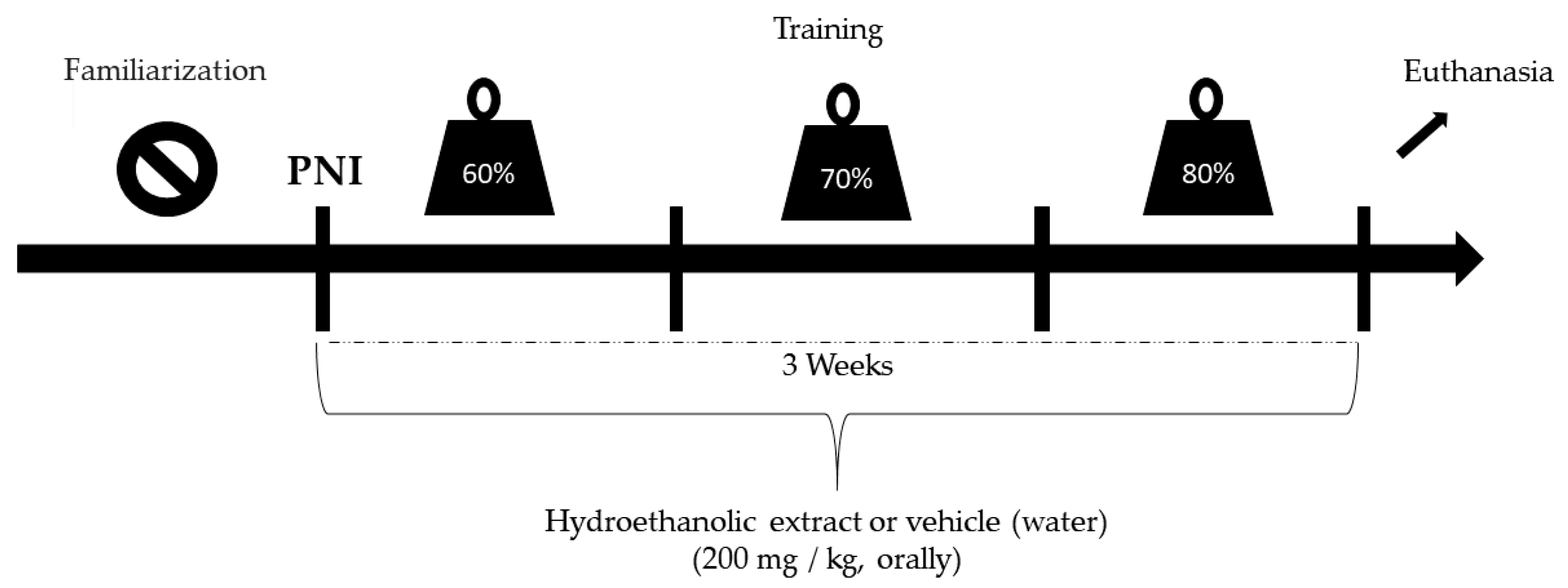
2.1. PNI Induction
2.2. Application of Therapeutic Resources
2.3. RT
2.4. Collection and Extraction of the Barks of B.virgilioides
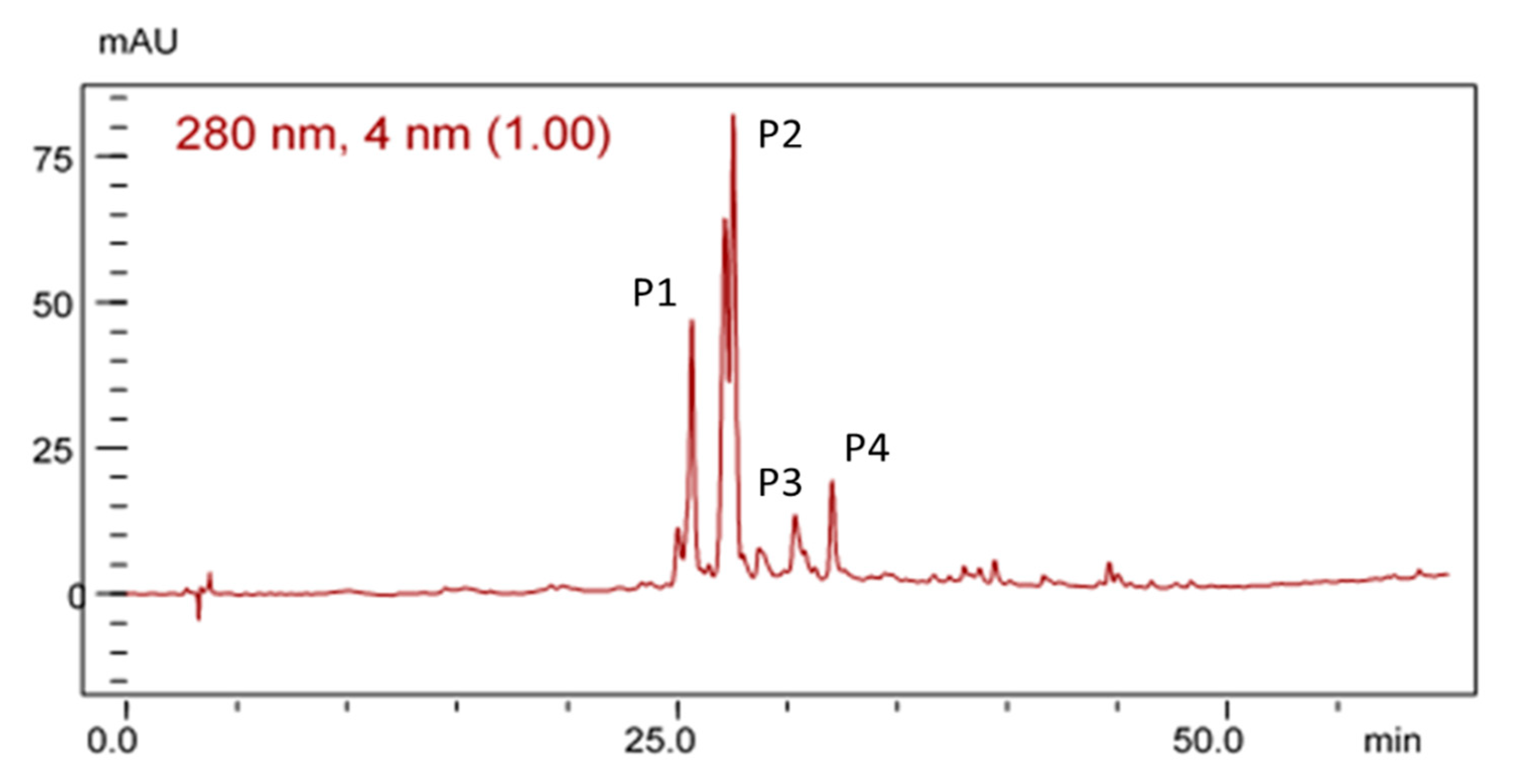
2.5. Phytochemical Prospecting
2.6. Quantification of Total Phenol Content (PC)
2.7. Total Phenols
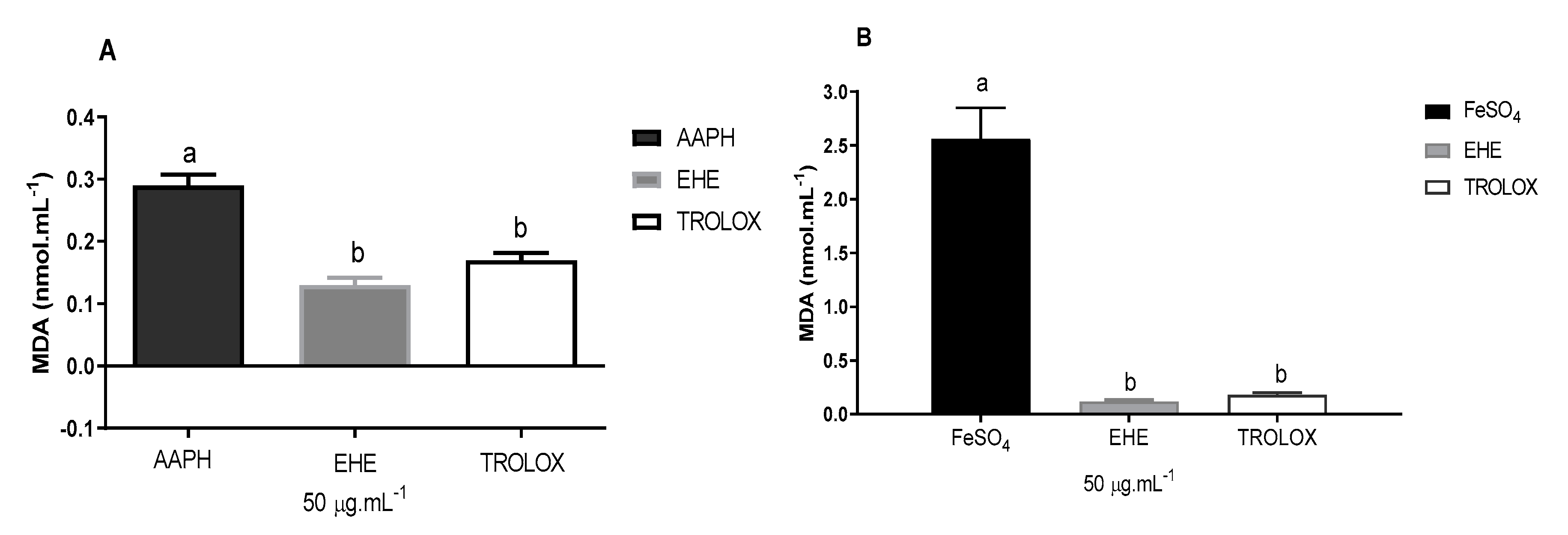
2.8. DPPH Free Radical Scavenging Activity
2.9. Euthanasia and Collection of Biological Material
2.10. Oxidative Stress (OS) Analysis
2.10.1. Determination of Malondialdehyde/Thiobarbituric Acid (TBARS) Reactive Substances In Vivo
2.10.2. Determination of Total Sulfhydryls (Thiols)
2.11. Statistical Analyses
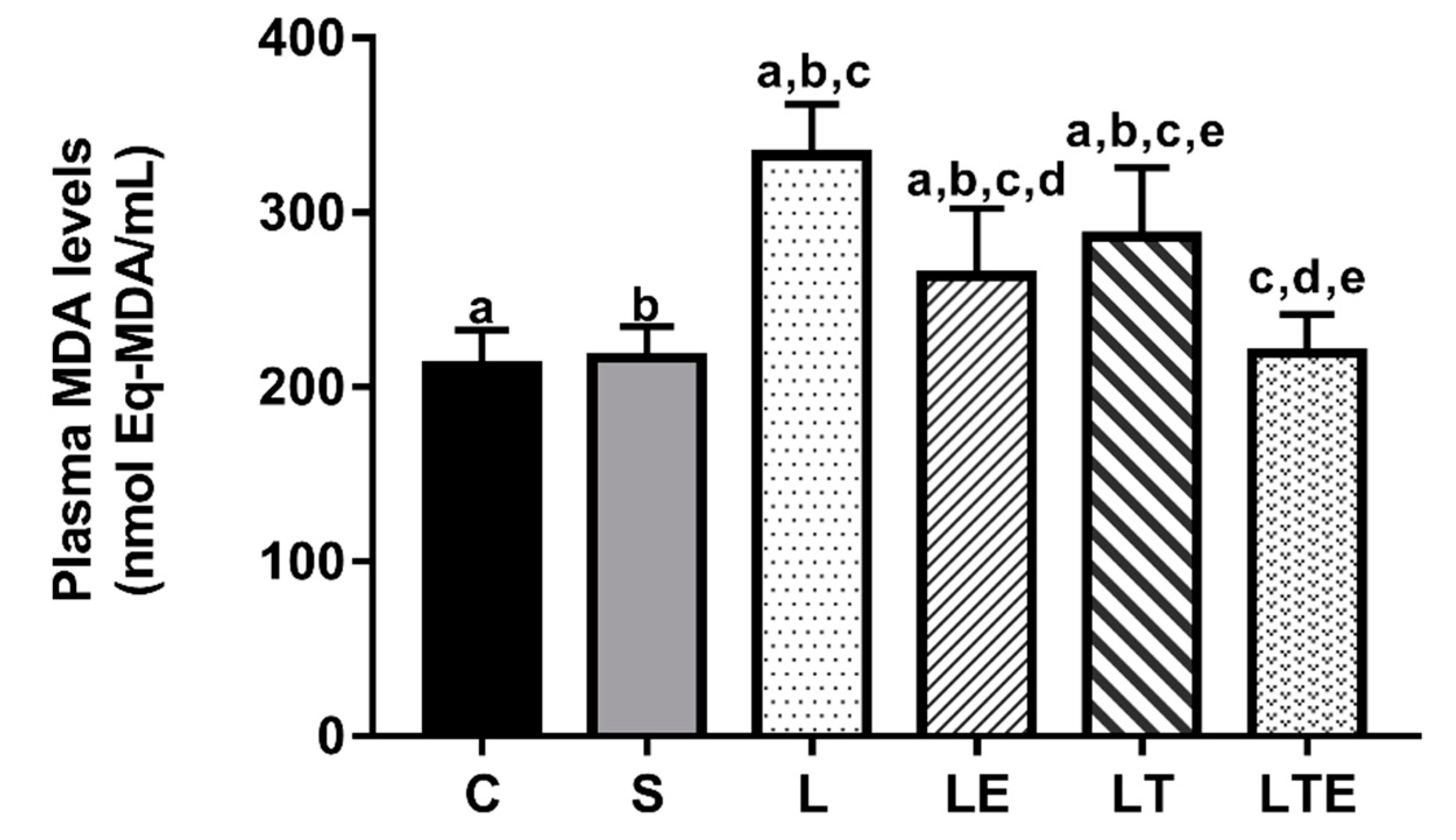
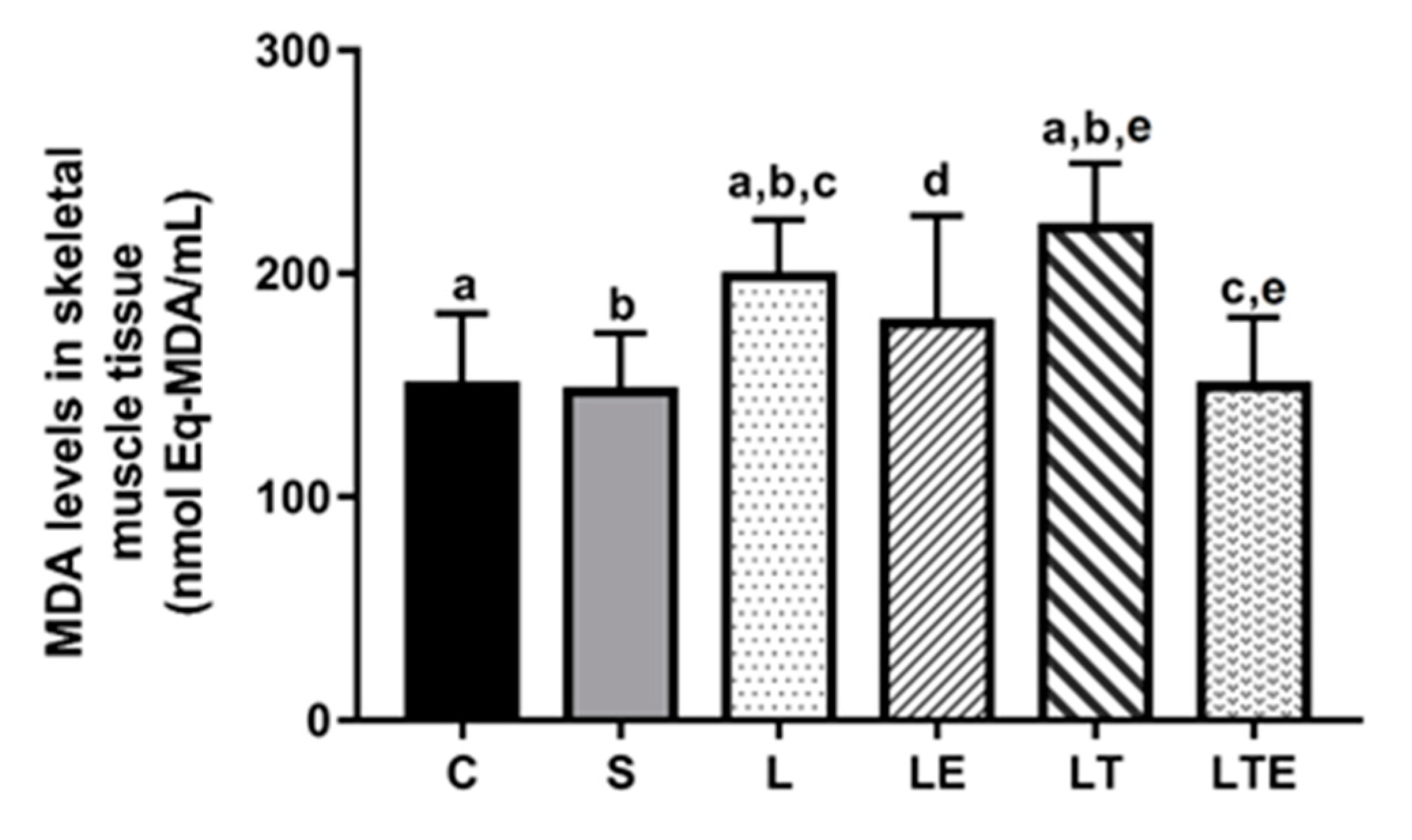
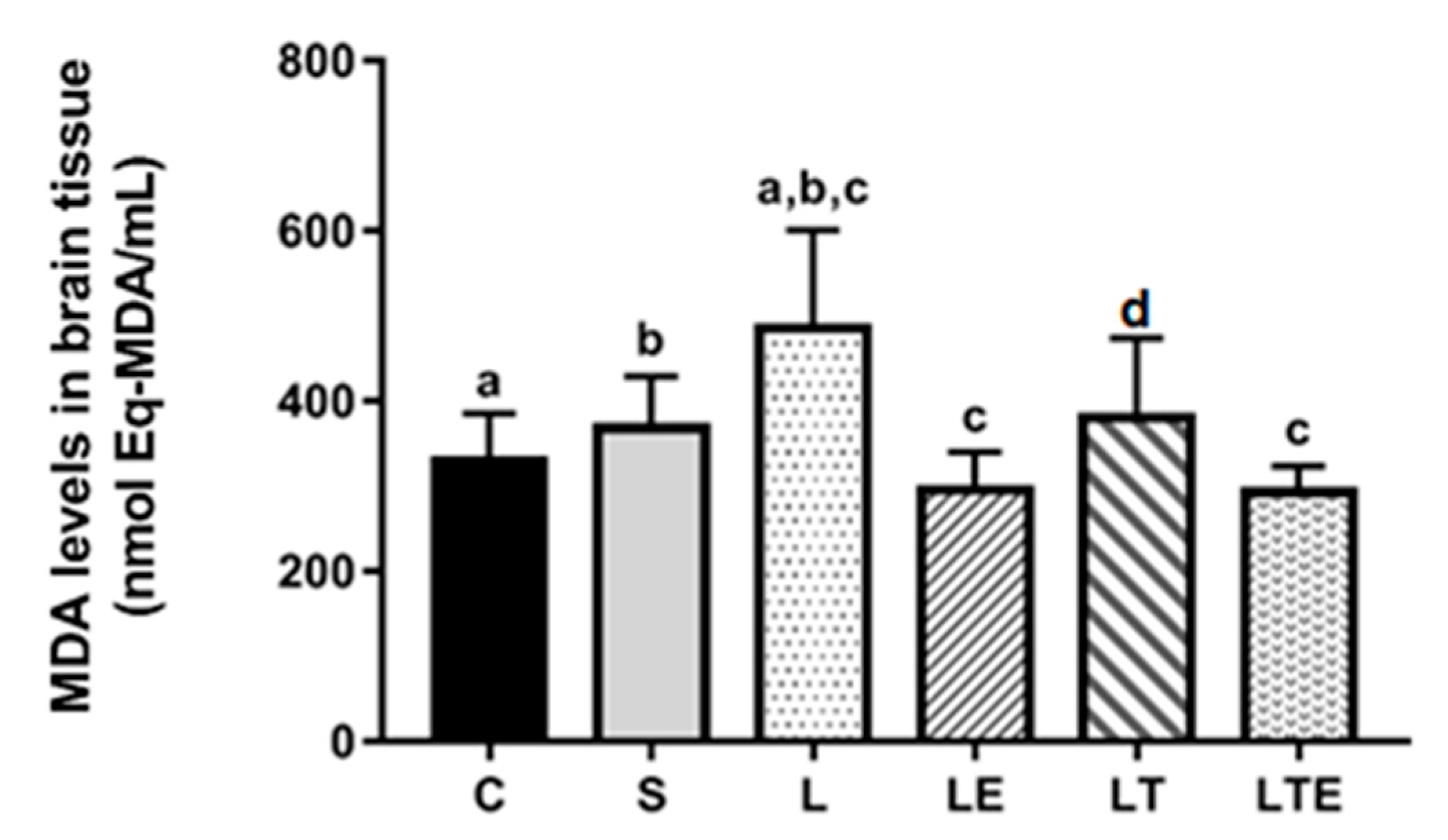
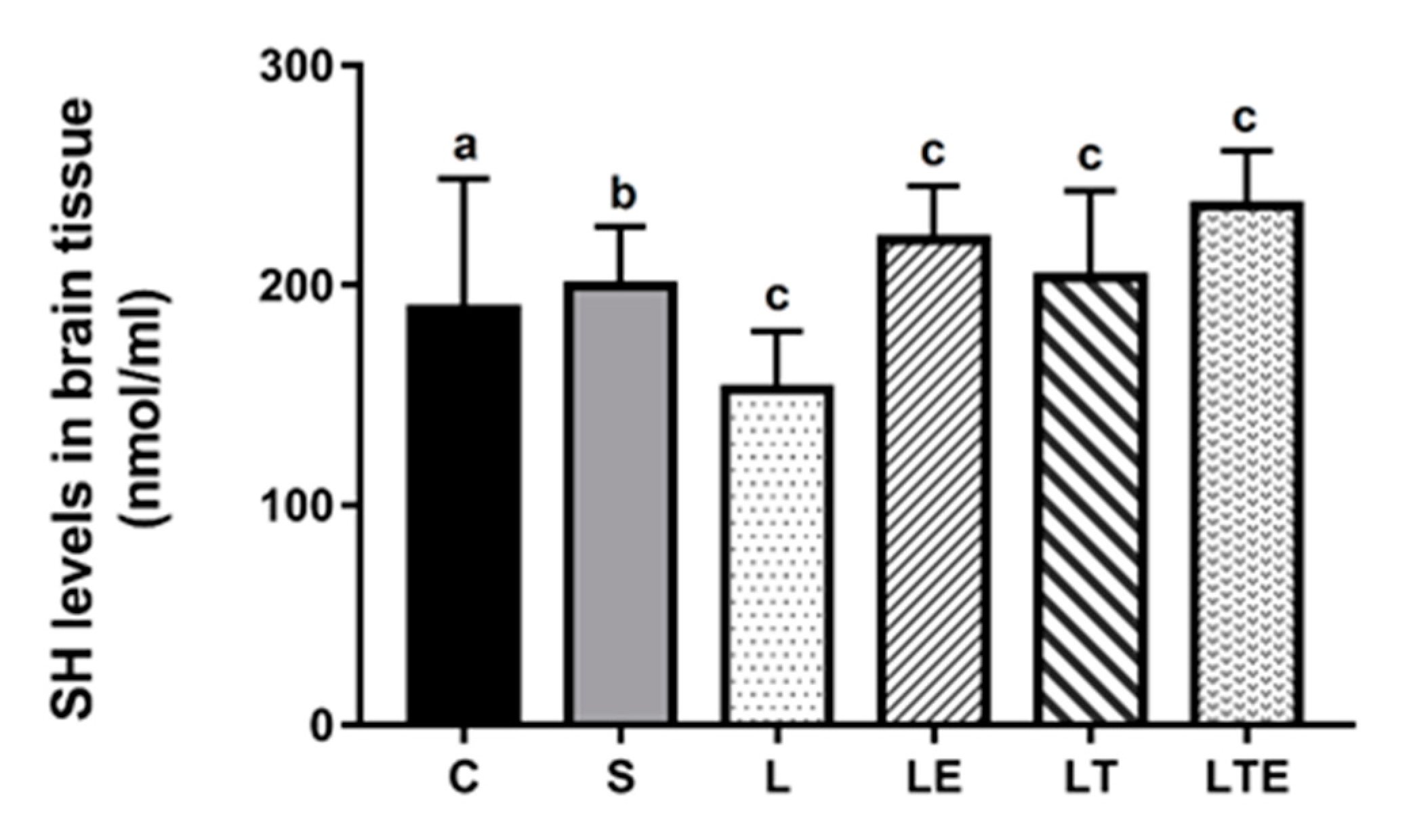
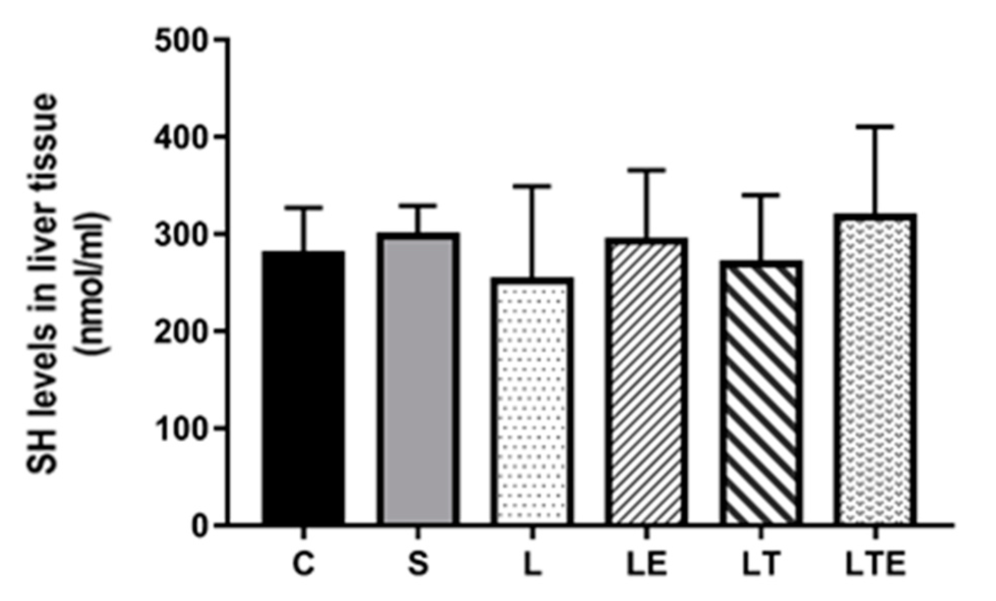
3. Results
4. Discussion
5. Conclusions
Author Contributions
Funding
Acknowledgments
Conflicts of Interest
References
- Wang, E.; Inaba, K.; Byerly, S.; Escamilla, D.; Cho, J.; Carey, J.; Stevanovic, M.; Ghiassi, A.; Demetriades, D. Optimal timing for repair of peripheral nerve injuries. J. Trauma Acute Care Surg. 2017, 83, 875–881. [Google Scholar]
- Saadat, S.; Eslami, V.; Rahimi-Movaghar, V. The incidence of peripheral nerve injury in trauma patients in Iran. Turk. J. Trauma Emerg. Surg. 2011, 17, 539–544. [Google Scholar]
- Huckhagel, T.; Nüchtern, J.; Regelsberger, J.; Lefering, R. Trauma DGU. Nerve injury in severe trauma with upper extremity involvement: Evaluation of 49,382 patients from the Trauma Register DGU® between 2002 and 2015 Scand. J. Trauma, Resus. Emerg. Med. 2018, 26, 76. [Google Scholar]
- Kouyoumdjian, J.A.; Graça, C.R.; Ferreira, V.F.M. Peripheral nerve injuries: A retrospective survey of 1124 cases. Neurol. India 2017, 65, 551–555. [Google Scholar]
- Ding, Z.; Cao, J.; Shen, Y.; Zou, Y.; Yang, X.; Zhou, W.; Guo, Q.; Huang, C. Resveratrol promotes nerve regeneration via activation of p300 acetyltransferase-mediated VEGF signaling in a rat model of sciatic nerve crush injury. Front. Neurosci 2018, 12, 341. [Google Scholar] [PubMed]
- Goswami, R.; Anastakis, D.J.; Katz, J.; Davis, K.D. A longitudinal study of pain, personality, and brain plasticity following peripheral nerve injury. Pain 2016, 157, 729–739. [Google Scholar] [CrossRef] [PubMed]
- Rivera, J.C.; Glebus, G.P.; Cho, M.S. Disability following combat-sustained nerve injury of the upper limb. Bone Joint J. 2014, 96, 254–258. [Google Scholar] [CrossRef]
- Gray, B. Quality of life following traumatic brachial plexus injury: A questionnaire study. Int. J. Orthop Trauma Nurs. 2016, 22, 29–35. [Google Scholar]
- Miller, C.; Peek, A.L.; Power, D.; Heneghan, N.R. Psychological consequences of traumatic upper limb peripheral nerve injury: A systematic review. Hand Ther. 2017, 22, 35–45. [Google Scholar]
- Eser, F.; Aktekin, L.A.; Bodur, H.; Atan, C. Etiological factors of traumatic peripheral nerve injuries. Neurol. India 2009, 57, 434–437. [Google Scholar]
- Safakhah, H.A.; Moradi Kor, N.; Bazargani, A.; Bandegi, A.R.; Gholami Pourbadie, H.; Khoshkholgh-Sima, B.; Ghanbari, A. Forced exercise attenuates neuropathic pain in chronic constriction injury of male rat: An investigation of oxidative stress and inflammation. J. Pain Res. 2017, 10, 1457–1466. [Google Scholar] [CrossRef] [PubMed] [Green Version]
- Vijayavenkataraman, S. Nerve guide conduits for peripheral nerve injury repair: A review on design, materials and fabrication methods. Acta Biomater. 2020, 106, 54–69. [Google Scholar] [CrossRef]
- Ferrante, M.A. The assessment and management of peripheral nerve trauma. Curr. Treat. Options Neurol. 2018, 20, 25. [Google Scholar] [CrossRef] [PubMed]
- Kubiak, C.A.; Kung, T.A.; Brown, D.L.; Cederna, P.S.; Kemp, S.W.P. State-of-the-art techniques in treating peripheral nerve injury. Plast. Reconstr. Surg. 2018, 141, 702–710. [Google Scholar] [CrossRef]
- Lu, Y.; Li, R.; Zhu, J.; Wu, Y.; Li, D.; Dong, L.; Li, Y.; Wen, X.; Yu, F.; Zhang, H.; et al. Fibroblast growth factor 21 facilitates peripheral nerve regeneration through suppressing oxidative damage and autophagic cell death. J. Cell. Mol. Med. 2019, 23, 497–511. [Google Scholar] [CrossRef] [PubMed]
- Shen, Y.; Zhang, R.; Xu, L.; Wan, Q.; Zhu, J.; Gu, J.; Huang, Z.; Ma, W.; Shen, M.; Ding, F.; et al. Microarray analysis of gene expression provides new insights into denervation-induced skeletal muscle atrophy. Front. Physiol. 2019, 10, 1298. [Google Scholar] [CrossRef] [Green Version]
- Pisoschi, A.M.; Pop, A. The role of antioxidants in the chemistry of oxidative stress: A review. Eur. J. Med. Chem. 2015, 97, 55–74. [Google Scholar] [CrossRef]
- Oñate, M.; Catenaccio, A.; Martínez, G.; Armentano, D.; Parsons, G.; Kerr, B.; Hetz, C.; Court, F.A. Activation of the unfolded protein response promotes axonal regeneration after peripheral nerve injury. Sci. Rep. 2016, 6, 21709. [Google Scholar] [CrossRef] [Green Version]
- Sullivan, R.; Dailey, T.; Duncan, K.; Abel, N.; Borlongan, C.V. Peripheral nerve injury: Stem cell therapy and peripheral nerve transfer. Int. J. Mol. Sci. 2016, 17, 2101. [Google Scholar] [CrossRef]
- Martinez de Albornoz, P.M.; Delgado, P.J.; Forriol, F.; Maffulli, N. Non-surgical therapies for peripheral nerve injury. Br. Med. Bull. 2011, 100, 73–100. [Google Scholar] [CrossRef] [Green Version]
- Gröpel, P.; Urner, M.; Pruessner, J.C.; Quirin, M. Endurance-and resistance-trained men exhibit lower cardiovascular responses to psychosocial stress than untrained men. Front. Psychol. 2018, 9, 852. [Google Scholar] [PubMed] [Green Version]
- Stavres, J.; Zeigler, M.P.; Bayles, P.M. Six weeks of moderate functional resistance training increases basal metabolic rate in sedentary adult women. Int. J. Exer. Sci. 2018, 11, 32–41. [Google Scholar]
- De Sousa, E.C.; Abrahin, O.; Ferreira, A.L.L.; Rodrigues, R.P.; Alves, E.A.C.; Vieira, R.P. Resistance training alone reduces systolic and diastolic blood pressure in prehypertensive and hypertensive individuals: Meta-analysis. Hypertens. Res. 2017, 40, 927–931. [Google Scholar] [PubMed]
- Lovison, K.; Vieira, L.; Kunz, R.I.; Da Silva Scarton, S.R.; Antunes, J.S.; Karvat, J.; Peretti, A.L.; Bertolini, G.R.F.; Brancalhão, R.M.C.; Leme Beu, C.C.; et al. Resistance exercise recovery morphology and AQP1 expression in denervated soleus muscle of Wistar rats. Motricidade 2018, 14, 40–50. [Google Scholar]
- Araújo-Filho, H.G.; Quintans-Júnior, L.J.; Barreto, A.S.; Almeida, J.R.; Barreto, R.S.; Quintans, J.S. Neuroprotective effect of natural products on peripheral nerve degeneration: A systematic review. Neurochem. Res. 2016, 41, 647–658. [Google Scholar]
- Silva, T.L.; Fernandes, J.B.; Silva, M.; Consolaro, H.N.; de Sousa, L.R.; Vieira, P.C. New cathepsin V inhibitor from stems of Bowdichia virgilioides. Rev. Bras. Farmacogn. 2019, 29, 491–494. [Google Scholar]
- dos Santos, J.L.; Dantas, R.E.A.; Lima, C.A.; de Araújo, S.S.; de Almeida, E.C.V.; Marçal, A.C.; dos Santos Estevam, C. Protective effect of a hydroethanolic extract from Bowdichiavirgilioides on muscular damage and oxidative stress caused by strenuous resistance training in rats. J. Int. Soc. Sports Nutr. 2014, 11, 58. [Google Scholar]
- Malanotte, J.A.; Kakihata, C.M.M.; Karvat, J.; Brancalhão, R.M.C.; Ribeiro, L.F.C.; Bertolini, G.R.F. Jumping in aquatic environment after sciatic nerve compression: Nociceptive evaluation and morphological characteristics of the soleus muscle of Wistar rats. Einstein 2017, 15, 77–84. [Google Scholar]
- Hornberger, T.A., Jr.; Farrar, R.P. Physiological hypertrophy of the FHL muscle following 8 weeks of progressive resistance exercise in the rat. Can. J. Appl. Physiol. 2004, 29, 16–31. [Google Scholar] [CrossRef]
- Matos, F.J.A. Introduction to Experimental Phytochemistry, 3rd ed.; Editora UFC: Fortaleza, Brazil, 2009; p. 148. [Google Scholar]
- Sousa, C.M.d.M.; Vieira-Junior, G.M.; Ayres, M.C.C.; Costa, C.L.S.d.; Araújo, D.S.; Cavalcante, L.C.D.; Barros, E.D.S.; Araújo, P.B.d.M.; Brandão, M.S.; Chaves, M.H. Total phenols and antioxidant activity of five medicinal plants. Quím Nova. 2007, 30, 351–355. [Google Scholar] [CrossRef]
- Brand-Williams, W.; Cuvelier, M.E.; Berset, C. Use of a free radical method to evaluate antioxidant activity. LWT Food Sci. Technol. 1995, 28, 25–30. [Google Scholar] [CrossRef]
- Scherer, R.; Godoy, H.T. Antioxidant activity index (AAI) by the 2,2-diphenyl-1-picrylhydrazyl method. Food Chem. 2009, 112, 654–658. [Google Scholar] [CrossRef]
- Lapenna, D.; Ciofani, G.; Pierdomenico, S.D.; Giamberardino, M.A.; Cuccurullo, F. Reaction conditions affecting the relationship between thiobarbituric acid reactivity and lipid peroxidesin human plasma. Free Radic. Biol. Med. 2001, 31, 331–335. [Google Scholar] [CrossRef]
- Faure, P.; Lafond, J.L. Measurement of plasma sulfhydryl and carbonyl groups as a possible indicator of protein oxidation. In Analysis of Free Radicals in Biological Systems; Birkhäuser: Basel, Switzerland, 1995; pp. 237–248. [Google Scholar]
- Di Meo, S.; Napolitano, G.; Venditti, P. Mediators of physical activity protection against ROS-linked skeletal muscle damage. Int. J. Mol. Sci. 2019, 20, 3024. [Google Scholar] [CrossRef] [PubMed] [Green Version]
- Khoobkhahi, N.; Delavar, R.; Nayebifar, S.H. The combinatory effects of combined training (endurance–resistance) and garlic supplementation on oxidative stress and antioxidant adaptations in untrained boys. Sci. Sports 2019, 34, 410.e1–410.e7. [Google Scholar] [CrossRef]
- Lang, F.; Aravamudhan, S.; Nolte, H.; Türk, C.; Hölper, S.; Müller, S.; Günther, S.; Blaauw, B.; Braun, T.; Krüger, M. Dynamic changes in the mouse skeletal muscle proteome during denervation-induced atrophy. Dis. Models Mech. 2017, 10, 881–896. [Google Scholar] [CrossRef] [Green Version]
- Possamai, F.; Siepko, C.M.; André, E.S. Investigation of the effects of therapeutic exercise with peripheral nerve regeneration. Acta Fisiátrica 2010, 17, 142–147. [Google Scholar]
- Qiu, J.; Yang, X.; Wang, L.; Zhang, Q.; Ma, W.; Huang, Z.; Bao, Y.; Zhong, L.; Sun, H.; Ding, F. Isoquercitrin promotes peripheral nerve regeneration through inhibiting oxidative stress following sciatic crush injury in mice. Ann. Transl. Med. 2019, 7, 680. [Google Scholar] [CrossRef]
- Tsikas, D. Assessment of lipid peroxidation by measuring malondialdehyde (MDA) and relatives in biological samples: Analytical and biological challenges. Anal. Biochem. 2017, 524, 13–30. [Google Scholar] [CrossRef]
- Garcia, S.C.; Grotto, D.; Bulcão, R.P.; Moro, A.M.; Roehrs, M.; Valentini, J.; de Freitas, F.A.; Paniz, C.; Bubols, G.B.; Charão, M.F. Evaluation of lipid damage related to pathological and physiological conditions. Drug Chem. Toxicol. 2013, 36, 306–312. [Google Scholar] [CrossRef]
- Uslusoy, F.; Nazıroğlu, M.; Övey, İ.S.; Sönmez, T.T. Hypericum perforatum L. supplementation protects sciatic nerve injury-induced apoptotic, inflammatory and oxidative damage to muscle, blood and brain in rats. J. Pharm. Pharmacol. 2019, 71, 83–92. [Google Scholar] [CrossRef] [PubMed] [Green Version]
- Chen, Y.W.; Chiu, C.C.; Hsieh, P.L.; Hung, C.H.; Wang, J.J. Treadmill training combined with insulin suppresses diabetic nerve pain and cytokines in rat sciatic nerve. Anesth. Analg. 2015, 121, 239–246. [Google Scholar] [CrossRef] [PubMed]
- Chris, I.C.; Clichici, A.; Nagy, A.L.; Oros, A.; Catoi, C.; Clichici, S. Quercetin in association with moderate exercise training attenuates injuries induced by experimental diabetes in sciatic nerves. J. Physiol. Pharmacol. 2017, 68, 877–886. [Google Scholar]
- Brown, D.R.; Gough, L.A.; Deb, S.K.; Sparks, S.A.; McNaughton, L.R. Astaxanthin in exercise metabolism, performance and recovery: A review. Front. Nutr. 2017, 4, 76. [Google Scholar] [CrossRef] [Green Version]
- Steinbacher, P.; Eckl, P. Impact of oxidative stress on exercising skeletal muscle. Biomolecules 2015, 5, 356–377. [Google Scholar] [CrossRef]
- He, F.; Li, J.; Liu, Z.; Chuang, C.C.; Yang, W.; Zuo, L. Redox mechanism of reactive oxygen species in exercise. Front. Physiol. 2016, 7, 486. [Google Scholar] [CrossRef] [Green Version]
- Vargas-Mendoza, N.; Morales-González, Á.; Madrigal-Santillán, E.O.; Madrigal-Bujaidar, E.; Álvarez-González, I.; García-Melo, L.F.; Anguiano-Robledo, L.; Fregoso-Aguilar, T.; Morales-Gonzalez, J.A. Antioxidant and adaptative response mediated by Nrf2 during physical exercise. Antioxidants 2019, 8, 196. [Google Scholar] [CrossRef] [Green Version]
- Park, S.Y.; Kwak, Y.S. Impact of aerobic and anaerobic exercise training on oxidative stress and antioxidant defense in athletes. J. Exerc. Rehabil. 2016, 12, 113–117. [Google Scholar] [CrossRef]
- Nemati, Z.; Arazi, H.; Sariri, R. Salivary antioxidants status following two resistance exercise systems in young women athletes. Sports Med. J. 2017, 13, 2911–2917. [Google Scholar]
- Padilha, C.S.; Ribeiro, A.S.; Fleck, S.J.; Nascimento, M.A.; Pina, F.L.; Okino, A.M.; Venturini, D.; Barbosa, D.S.; Mayhew, J.L.; Cyrino, E.S. Effect of resistance training with different frequencies and detraining on muscular strength and oxidative stress biomarkers in older women. Age 2015, 37, 104. [Google Scholar] [CrossRef]
- Zhao, J.; Luo, D.; Liang, Z.; Lao, L.; Rong, J. Plant natural product puerarin ameliorates depressive behaviors and chronic pain in mice with spared nerve injury (SNI). Mol. Neurobiol. 2017, 54, 2801–2812. [Google Scholar]
- Barbosa, R.A.; Nunes, T.L.; Nunes, T.L.; da Paixão, A.O.; Belo Neto, R.; Moura, S.; Albuquerque Junior, R.L.; Cândido, E.A.; Padilha, F.F.; Quintans-Júnior, L.J.; et al. Hydroalcoholic extract of red propolis promotes functional recovery and axon repair after sciatic nerve injury in rats. Pharm. Biol. 2016, 54, 993–1004. [Google Scholar] [PubMed] [Green Version]
- Hsu, C.C.; Huang, H.C.; Wu, P.T.; Tai, T.W.; Jou, I.M. Sesame oil improves functional recovery by attenuating nerve oxidative stress in a mouse model of acute peripheral nerve injury: Role of Nrf-2. J. Nutr. Biochem. 2016, 38, 102–106. [Google Scholar] [PubMed]
- dos Santos Reis, M.D.; de Ara&ujo Vieira, L.F.; Brandão, A.R.E.A.; Barreto, E.; Smaniotto, S. The effects of aqueous extract of Bowdichia virgilioides Kunth in the immune functions of thymocytes and B-lymphocytes. J. Med. Plants Res. 2020, 14, 247–257. [Google Scholar]
- Assis, I.B.; da Rocha, E.M.M.; Guimarães, D.S.M.; do Nascimento Pereira, G.A.; Pereira, F.P.; Ferreira, J.M.S.; Barreto, E.O. Evaluation of biological activity, toxicity, and phytochemical content of Bowdichiavirgilioides (Fabaceae) aqueous extract. Pharmacogn. J. 2018, 14, 403. [Google Scholar]
- Barros, W.M.; Rao, V.S.N.; Silva, R.M.; Lima, J.C.S.; Martins, D.T.O. Anti-inflammatory effect of the ethanolic extract from Bowdichia virgilioides H.B.K stem bark. An. Acad. Bras. Cienc. 2010, 82, 609–616. [Google Scholar]
- Thomazzi, S.M.; Silva, C.B.; Silveira, D.C.R.; Vasconcellos, C.L.C.; Lira, A.F.; Cambui, E.V.F.; Estevam, C.S.; Antoniolli, A.R. Antinociceptive and anti-inflammatory activities of Bowdichia virgilioides (sucupira). J. Ethnopharmacol. 2010, 127, 451–456. [Google Scholar]
- Shan, A.Y.K.V.; de Almeida, E.C.V.; dos Santos, A.E.L.L.M.; de Lima, A.D.C.B.; de Souza Santos, C.C.; de Santana Souza, M.I.T.; Damascena, N.P.; de Araújo, S.S.; dos Santos, J.L.; Paixão, M.S.; et al. Antioxidant and antinociceptive effect of the hydroethanolic extract and fractions of the bark of Bowdichia virgilioides in orofacial pain. Afr. J. Pharm. Pharmacol. 2016, 10, 320–329. [Google Scholar]
- Vieira, L.F.d.A.; Reis, M.D.d.S.; Brandão, A.R.A.; Viana, I.M.M.N.; da Silva, J.P.; Barreto, E.; Smaniotto, S. Anxiolytic-like effect of the extract from Bowdichiavirgilioides in mice. Rev. Bras. Farmacognosia 2013, 23, 680–686. [Google Scholar]
- Sarubbo, F.; Esteban, S.; Miralles, A.; Moranta, D. Effects of resveratrol and other polyphenols on Sirt1: Relevance to brain function during aging. Curr. Neuropharmacol. 2018, 16, 126–136. [Google Scholar]
- Yahfoufi, N.; Alsadi, N.; Jambi, M.; Matar, C. The immunomodulatory and anti-inflammatory role of polyphenols. Nutrients 2018, 10, 1618. [Google Scholar] [CrossRef] [PubMed] [Green Version]
- Ullah, I.; Choe, Y.H.; Khan, M.; Bharti, D.; Shivakumar, S.B.; Lee, H.J.; Son, Y.B.; Shin, Y.; Lee, S.L.; Park, B.W.; et al. Dental pulp-derived stem cells can counterbalance peripheral nerve injury-induced oxidative stress and supraspinal neuro-inflammation in rat brain. Sci. Rep. 2018, 8, 15795. [Google Scholar] [CrossRef]
- Pinho, R.A.; Aguiar, A.S.; Radák, Z. Effects of resistance exercise on cerebral redox regulation and cognition: An interplay between muscle and brain. Antioxidants 2019, 8, 529. [Google Scholar] [CrossRef] [PubMed] [Green Version]
- Filosa, S.; Di Meo, F.; Crispi, S. Polyphenols-gut microbiota interplay and brain neuromodulation. Neural Regen. Res. 2018, 13, 2055–2059. [Google Scholar]
- Zhang, L.; Wang, X.; Cueto, R.; Effi, C.; Zhang, Y.; Tan, H.; Qin, X.; Ji, Y.; Yang, X.; Wang, H. Biochemical basis and metabolic interplay of redox regulation. Redox Biol. 2019, 26, 101284. [Google Scholar] [CrossRef] [PubMed]
- McLeay, Y.; Stannard, S.; Houltham, S.; Starck, C. Dietary thiols in exercise: Oxidative stress defence, exercise performance, and adaptation. J. Int. Soc. Sports Nutr. 2017, 14, 12. [Google Scholar] [CrossRef] [Green Version]


| Components | EHE |
|---|---|
| Phenols | + |
| Tannins | + |
| Flavonoids | + |
| Xanthones | + |
| Categories | + |
| Pentacyclic triterpenoids and free steroids | + |
| Saponins | − |
| Alkaloids | + |
| Sample | EC50(µg/mL) | PI (%) * | AAI ** |
|---|---|---|---|
| EHE | 29.50 ± 2.40 a | 42.90 | 0.84 |
| GallicAcid | 1.05 ± 0.20 a | 92.06 | 23.80 |
© 2020 by the authors. Licensee MDPI, Basel, Switzerland. This article is an open access article distributed under the terms and conditions of the Creative Commons Attribution (CC BY) license (http://creativecommons.org/licenses/by/4.0/).
Share and Cite
Costa, L.S.; Aidar, F.J.; Matos, D.G.d.; Oliveira, J.U.d.; Santos, J.L.d.; Almeida-Neto, P.F.d.; Souza, R.F.d.; Pereira, D.D.; Garrido, N.D.; Nunes-Silva, A.; et al. Effects of Resistance Training and Bowdichia virgilioides Hydroethanolic Extract on Oxidative Stress Markers in Rats Submitted to Peripheral Nerve Injury. Antioxidants 2020, 9, 941. https://doi.org/10.3390/antiox9100941
Costa LS, Aidar FJ, Matos DGd, Oliveira JUd, Santos JLd, Almeida-Neto PFd, Souza RFd, Pereira DD, Garrido ND, Nunes-Silva A, et al. Effects of Resistance Training and Bowdichia virgilioides Hydroethanolic Extract on Oxidative Stress Markers in Rats Submitted to Peripheral Nerve Injury. Antioxidants. 2020; 9(10):941. https://doi.org/10.3390/antiox9100941
Chicago/Turabian StyleCosta, Luana Santos, Felipe J. Aidar, Dihogo Gama de Matos, José Uilien de Oliveira, Jymmys Lopes dos Santos, Paulo Francisco de Almeida-Neto, Raphael Fabrício de Souza, Danielle Dutra Pereira, Nuno Domingos Garrido, Albená Nunes-Silva, and et al. 2020. "Effects of Resistance Training and Bowdichia virgilioides Hydroethanolic Extract on Oxidative Stress Markers in Rats Submitted to Peripheral Nerve Injury" Antioxidants 9, no. 10: 941. https://doi.org/10.3390/antiox9100941
APA StyleCosta, L. S., Aidar, F. J., Matos, D. G. d., Oliveira, J. U. d., Santos, J. L. d., Almeida-Neto, P. F. d., Souza, R. F. d., Pereira, D. D., Garrido, N. D., Nunes-Silva, A., Marçal, A. C., Estevam, C. d. S., Cabral, B. G. d. A. T., Reis, V. M., & Teixeira, M. M. (2020). Effects of Resistance Training and Bowdichia virgilioides Hydroethanolic Extract on Oxidative Stress Markers in Rats Submitted to Peripheral Nerve Injury. Antioxidants, 9(10), 941. https://doi.org/10.3390/antiox9100941